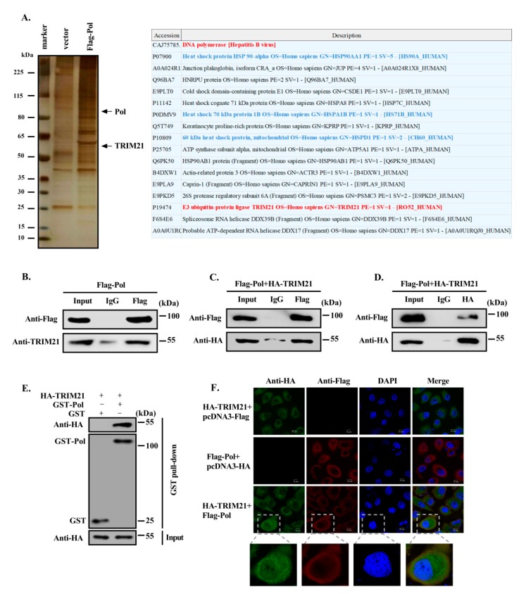
Figure 1.
TRIM21 interacts with HBV DNA Pol. (A) Huh7 cells were transfected with FLAG-HBV DNA Pol and vector control, and FLAG affinity beads were used to precipitate all proteins that might interact with HBV DNA Pol. The interacting proteins of HBV DNA Pol were screened by mass spectrometry. Some potential interacting partners of HBV DNA Pol are listed. Red marks the target protein, blue marks proteins that have been reported to interact with HBV DNA Pol. Data are representative of two independent experiments. (B) The FLAG-HBV DNA Pol expression plasmid was transfected into Huh7 cells. After 36h, a co-IP assay was carried out with anti-FLAG antibody and control IgG antibody. HBV DNA Pol was then detected with anti-FLAG antibody, and anti-TRIM21 antibody was used to detect the endogenous expression of TRIM21 in Huh7 cells by Western blot. (C) Huh7 cells were cotransfected with FLAG-HBV DNA Pol and HA-TRIM21, and co-IP was performed as described in (B) to detect the expression of HBV DNA Pol and TRIM21. (D) Huh7 cells were transfected as described in (C), co-IP was performed with anti-HA antibody, and HBV DNA Pol and HA-TRIM21 expression were detected by Western blot. (E) GST-HBV DNA Pol and HA-TRIM21 were transfected into Huh7 cells, 36h after transfection, GST beads were used to pull down GST-Pol with its interacting proteins. HBV DNA Pol was detected with anti-GST antibody, and anti-HA antibody was used to detect the expression of TRIM21. (F) FLAG-HBV DNA Pol and HA-TRIM21 were transfected into Huh7 cells; 36h after transfection, the mouse anti-FLAG TRITC-conjugated monoclonal antibody and rabbit anti-HA FITC-conjugated polyclonal antibody were used to stain the cells, and the DAPI were used to stain the nuclei. Then, confocal microscopy images were collected and analyzed for the colocalization of HBV DNA Pol and TRIM21. The scale is 10 μm. Data are representative of three independent experiments with three replicates each.

