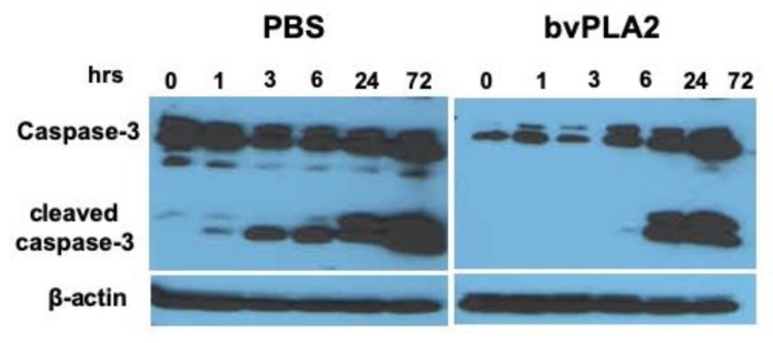
Figure 5.
Expression of caspase-3 in mouse splenocytes in the absence or presence of bvPLA2. Mouse splenocytes were treated with PBS or bvPLA2 and incubated at 37 °C for 0, 1, 3, 6, 24, and 72 h and then analyzed by western blotting with a monoclonal anti-caspase-3 antibody. Typical immunoblot of each sample showing the 32-kDa procaspase band and 12 and 17 kDa cleaved-caspase-3 bands. β-actin was used as a loading control for normalization. Data are representative of two individual experiments.

