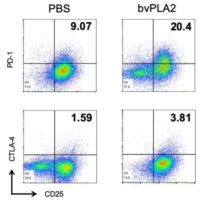
Figure 6.
Effect of bvPLA2 treatment on the levels of CTLA-4 and PD-1 in vitro. Flow cytometry of mouse splenocytes that were stimulated with anti-CD3/CD28 antibodies in the presence of bvPLA2. Representative dot-plots showed staining of CD25 and CTLA-4 or PD-1 expression. Live cells were gated with anti-CD4 antibody and subsequent gating based on expression of CD25 and CTLA-4 or PD-1 is depicted.

