Figure 3.
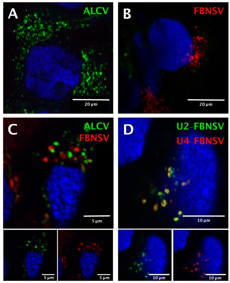
Localization of ALCV and FBNSV DNA in anterior midgut cells of A. craccivora. Co-labeling of ALCV (green) and FBNSV (8 segments probes, red) in aphids fed on plants infected with ALCV alone (A), FBNSV alone (B), or with both viruses (C). Co-labeling of the two FBNSV segments U2 (green) and U4 (red) in aphids fed on plants co-infected by FBNSV and ALCV (D). Top panels in C and D are images with merged color channels, and the corresponding split channels are at the bottom left (green) and right (red). Images A and B correspond to maximum intensity projections and images C and D to single optical sections. Cell nuclei are blue-stained with DAPI.

