Figure 7.
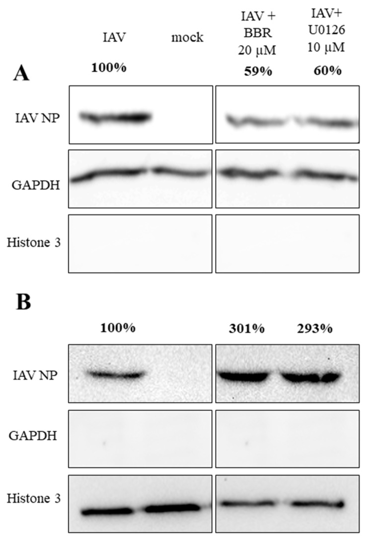
BBR treatment leads to the accumulation of influenza A virus NP in nuclei. The figure shows Western blot analyses of cytoplasmic (A) and nuclear (B) fractions of A549 cells infected with influenza A virus (TCID50 = 2000/mL) with the addition of DMSO solvent, U0126, or BBR at given concentrations. After 12 h, cytoplasmic and nuclear fractions of the cells were obtained and resolved by 12% SDS-PAGE. Influenza A nucleoprotein (IAV NP) was visualized by Western blot with anti-NP. GAPDH (glyceraldehyde 3-phosphate dehydrogenase) and histone 3 proteins were blotted as fraction purity markers. Percentage values show relative expression ratio of influenza A virus NP after normalization to GAPDH (A) or histone 3 (B) signal in each lane.

