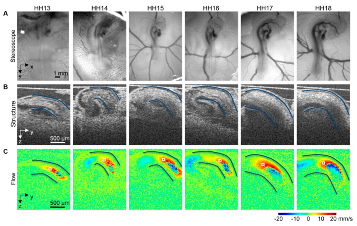
Figure 6.
Images of the avian heart over looping stages, HH13-HH18. (A) Optical images of chicken embryos in ovo on the top of the egg surface, (B) OCT structural two-dimensional longitudinal images of the heart outflow tract and neighboring structures and (C) corresponding Doppler OCT images. The heart outflow tract myocardial walls are outlined in (B,C), and an example corresponding point for velocity extraction and measurement is marked by a box in (C). Reproduced with permission from [173].

