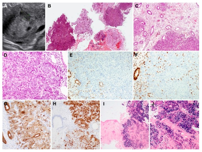
Figure 1.
Histopathological findings, immunostaining results, and Epstein–Barr virus-encoded RNA in situ hybridization (EBER-ISH) results of metastatic non-keratinizing undifferentiated carcinoma of the nasopharynx (case 1). (A) Abdominopelvic ultrasonography reveals a hypoechoic endometrial mass. (B) On scanning view, several endometrial tissue fragments are totally or partially replaced by tumor tissues. (C) Low-power magnification shows variable-sized tumor cell clusters infiltrating the endometrial stroma. The glands are not involved; however, they are surrounded by tumor cells. (D) High-power magnification exhibits an admixture of tumor cells showing syncytial growth and polymorphic inflammatory cells. The tumor cells possess large pleomorphic nuclei with conspicuous nucleoli. (E,F) Tumor cells are negative for (E) estrogen receptor (ER) or (F) progesterone receptor (PR); in contrast, these proteins are uniformly and strongly expressed in the nuclei of endometrial glandular epithelium and stromal cells. (G) Intensity of CK7 immunoreactivity is strong and moderate in the endometrial glandular epithelium and tumor cells, respectively. (H) Tumor cells display strong nuclear p63 expression. (I) EBER-ISH reveals positive signaling in tumor tissues (right). (J) Higher magnification of image (I) Tumor cells exhibit strongly positive nuclear staining. Original magnification: B: ×12.5; C: ×40; D: ×200; E–H: ×100, I: ×12.5; J: ×40.

