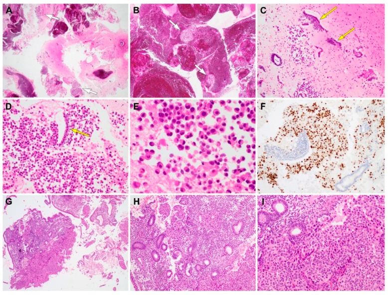
Figure 2.
Histopathological findings and immunostaining results of metastatic invasive lobular (case 2) and ductal (case 3) carcinoma. (A,B) On scanning view, the curetted specimen consists mainly of blood and fibrin, and a small amount of scattered tissue fragments (short white arrows). (C) Low-power magnification shows individual tumor cells and endometrial strips (long yellow arrows) dispersed in the background of a fibrinous exudate. (D) Medium-power magnification demonstrates a monotonous population of discohesive tumor cells. A long yellow arrow indicates the endometrial strip located between the tumor cells. (E) On high-power magnification, the tumor cells appear round to polygonal and possess eccentrically placed hyperchromatic nuclei and eosinophilic cytoplasm, morphologically compatible with invasive lobular carcinoma of the breast. (F) Tumor nuclei are strongly positive for GATA3, whereas the endometrial strips do not express GATA3. (G) Metastatic carcinoma cells infiltrating the endometrial stroma. (H) Presence of benign endometrial glands, which are entrapped within tumor cells clusters, favors the diagnosis of a metastatic lesion. (I) Cohesive tumor cells forming solid sheets and a few small glands, demonstrating morphological compatibility with invasive ductal carcinoma of the breast. They possess large pleomorphic nuclei and abundant eosinophilic cytoplasm. Original magnification: A and B: ×12.5; C: ×40; D: ×40; E: ×200; F: ×40; G: ×12.5; H: ×40, I: ×100.

