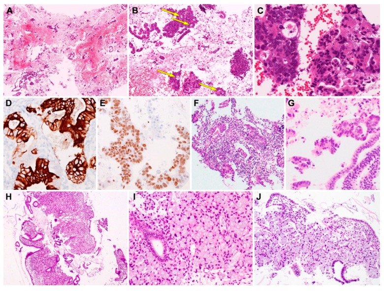
Figure 3.
Histopathological findings and immunostaining results of metastatic gastrointestinal carcinoma. Metastatic colorectal adenocarcinoma (case 4). (A) Curetted specimen consists mainly of blood, fibrin, and necrotic debris admixed with tumor tissue fragments of variable size. (B) Irregular-shaped clusters of tumor cells exhibit solid and cribriform architecture with small glandular lumina (long yellow arrows). (C) Intraluminal eosinophilic material, necrotic debris, and severe nuclear pleomorphism of the glandular epithelium are morphologically consistent with colorectal adenocarcinoma. (D) Tumor cells are strongly positive for CK20. (E) Tumor cell nuclei also react uniformly with CDX2. Metastatic colorectal adenocarcinoma (case 5). (F) In a few foci, infiltrating tumor tissues are associated with small amount of stromal desmoplasia and artifactual clefts. (G) Fragmented epithelium possess larger, more hyperchromatic tumor cell nuclei, compared with those of endometrial strips (right lower corner). Metastatic gastric (case 6) and appendiceal (case 7) signet ring cell carcinoma. (H) Metastatic signet ring cell carcinoma from the stomach involving the endometrial stroma. (I,J) Medium-power magnification demonstrating aggregates of signet ring cells with typical morphological features of large collections of intracytoplasmic mucin compressing the nuclei towards the periphery of the cell. The entrapped endometrial glands possess small and bland nuclei. The uninvolved endometrial stroma display bland-appearing spindle cells (right lower corner). Original magnification: A, ×12.5; B, ×40; C: ×200; D: ×200; E, ×200; F, ×100; G, ×200; H, ×40; I, ×200; J, ×100.

