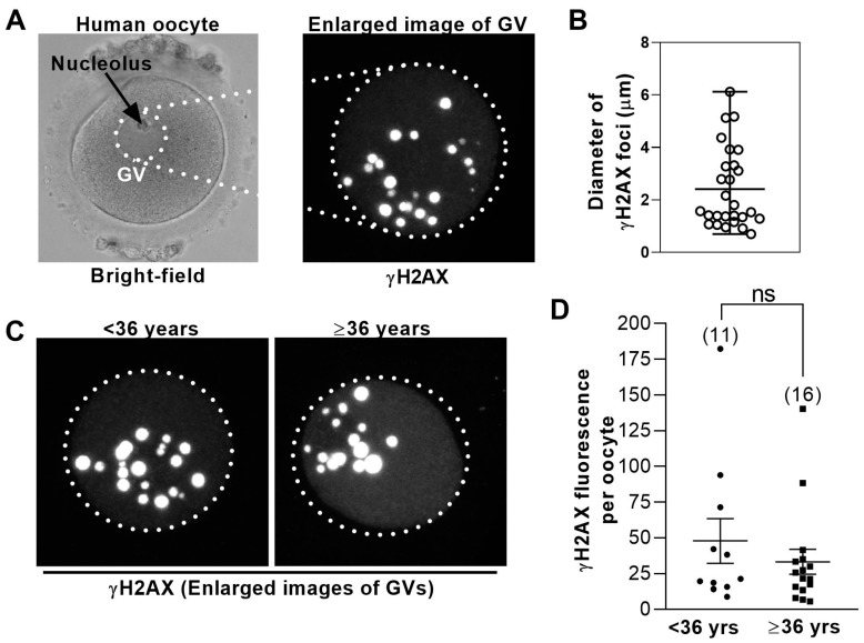
Figure 1.
Levels of DNA damage in human germinal vesicle (GV) oocytes are not age-dependent. (A) Shown is a bright-field image of a GV− stage human oocyte (left); dotted circle highlights the GV. To the right is an enlarged view of the GV immunostained for γH2AX. Shown is a maximum projection of multiple z-slices. (B) The sizes of γH2AX foci in human GVs vary widely. The diameters of individual γH2AX foci from the oocyte in (A) were measured using LAS software and plotted. Bars represent the mean and range. (C) Shown is the representative γH2AX immunostained GVs from younger (<36 years of age) and older (≥36 years of age) women. Images are maximum projections of multiple z-slices. (D) Total γH2AX fluorescence intensity within the GV was measured using confocal microscopy (see Methods) in oocytes from younger and older women (numbers of oocytes are shown in parenthesis) and plotted for each oocyte. Bars represent mean and SD.

