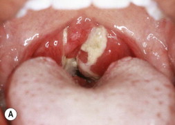
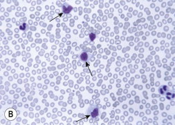
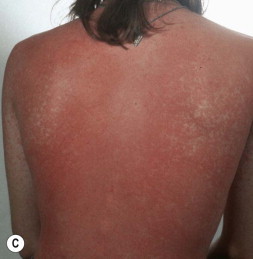
Figure 27-3.


(A) Pharyngeal erythema and exudate of Epstein–Barr virus (EBV). (B) Peripheral blood smear showing atypical lymphocytes (arrows) in a patient with EBV mononucleosis. Note the abundant cytoplasm with vacuoles, and deformation of cell by surrounding cells. (C) Diffuse erythematous raised rash in adolescent with EBV mononucleosis who received amoxicillin; note predominance on trunk and coalescence.
(Courtesy of J.H. Brien©.)
