
Figure 103-7.
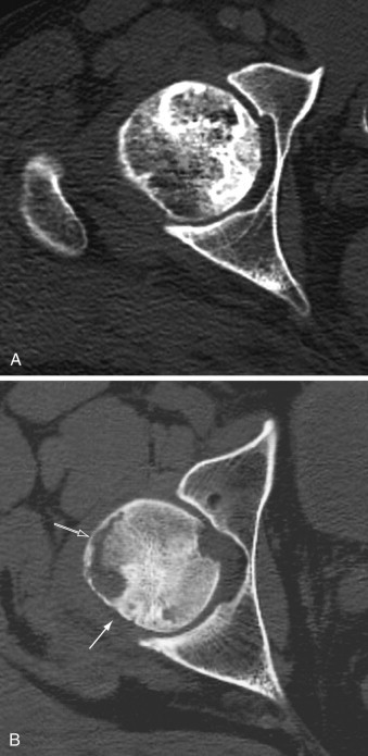
A, Computed tomography scan shows osteonecrosis of the femoral head. Although there are several sclerotic foci within the trabecular bone, the integrity of the osseous structures is preserved and the femoral head exhibits normal spherical shape. B, In more advanced stage of osteonecrosis of the femoral head, note increased sclerosis in the posterior aspect (solid arrow) and subchondral collapse of necrotic bone anterolaterally (open arrow).