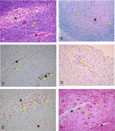
Fig. 35.2.

Acute disseminated encephalomyelitis. Photomicrographs showing a multiple sclerosis lesion with surrounding inflammatory response and loss of myelin. (A) Hematoxylin & eosin-luxol fast blue (H&E-LFB) stain showing perivascular loss of myelin (arrows showing myelin stained with LFB). (B) High magnification showing loss of myelin in the lesion center (asterisk) surrounded by intact myelin stained with LFB (arrows). (C and D) Stain for CD3 + T cells showing accumulation of T cells around the lesions (arrows). (E) CD68 stain showing macrophages surrounding blood vessels. (F) Glial fibrillary acidic protein (GFAP) stain showing reactive astrocytes (gliosis) surrounding blood vessels (arrows).
(Courtesy of Peter Pytel, MD, Department of Pathology, University of Chicago.)
