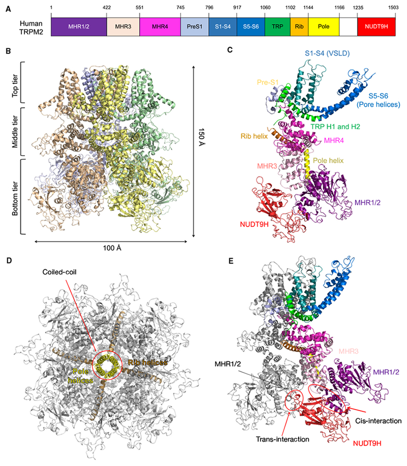
Fig. 1.

Apo-state structure of human TRPM2. (A) Domain arrangement of human TRPM2. (B) Ribbon diagram, dimensions, and three-tier architecture of human TRPM2. The four subunits are colored differently. (C) One subunit from the apo-state TRPM2 with domains colored individually. (D) The rib and pole helices form a central scaffold to support the TRPM2 channel. (E) The NUDT9H domain mediates extensive interactions with MHR domains both in cis and in trans.
