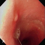
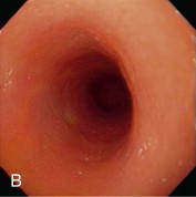
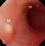
Figure 57-2.


Videoendoscopic appearance of the normal upper small intestine.
A, The major duodenal papilla (M) in the duodenum of the dog is the site of entry of the common bile duct and major pancreatic duct. The minor duodenal papilla (m) is seen in some but not all dogs distal to the major papilla and approximately 100 degrees clockwise from it. B, Normal descending duodenum in a dog; the distal flexure is visible in the distance. C, Peyer patches (lymphoid aggregates) in the duodenum appear as pale oval depressions along the antimesenteric border of the descending duodenum.
(Reprinted with permission from Lhermette P, Sobel D: BSAVA Manual of Canine and Feline Endoscopy and Endosurgery. Gloucester, UK, 2008, BSAVA Publications.)
