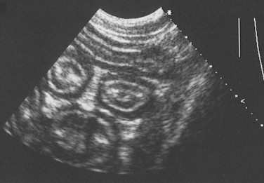
Figure 57-3.

Ultrasound Image of the Small Intestine.
Abdominal ultrasound image showing transverse image of three loops of bowel in a dog, with normal layering of the small intestinal wall.
(From Ettinger SJ and Feldman EC, editors: Textbook of Veterinary Internal Medicine, ed 7, Philadelphia. 2010, Saunders, p 1541.)
