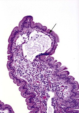
Figure 57-16.

A photomicrograph of a villi with lymphangiectasia. Note how the lacteal has engorged so much that now only the epithelium (arrow) is holding the chyle in the lacteal. This lacteal is about to rupture and release its contents into the intestinal lumen. It would be very easy to rupture this relatively fragile “balloon” if it were compressed, as might likely happen when using endoscopic biopsy forceps.
