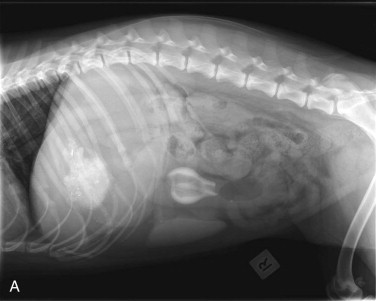
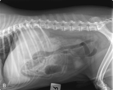
Figure 57-23.


A, Lateral abdominal radiograph of a dog with a radiopaque foreign body. Notice the fluid-distended loop of small intestine cranial to the object. Radiopaque sand-like material can be seen in the stomach. B, Ventrodorsal radiograph of the same dog. In the left caudal abdomen there is a focal region of increased soft tissue/mild mineral opacity within the intestinal lumen associated with fluid distention on one side, and gas distention (with fluid bubbles) on the other side.
