
eFigure 32-8.
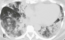
Seasonal influenza A infection progressing to respiratory failure with diffuse alveolar damage.
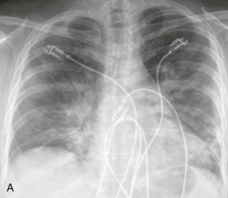
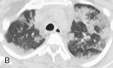
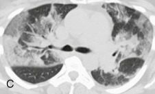
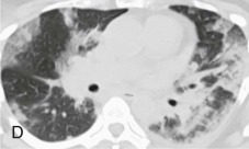
A, Frontal chest radiograph in a previously healthy 51-year-old woman with no significant previous medical history presenting to the emergency department with fever, cough, and nasal congestion shows multifocal, perihilar predominantly linear opacity and bronchovascular thickening and hazy opacities. The patient had been seen as an outpatient and treated with several broad-spectrum antibiotics with no improvement. At the time the chest radiograph was performed, the patient was mildly leukopenic with an oxygen saturation of 82% on room air. B–E, Axial chest CT displayed in lung windows shows multifocal, bilateral, nonsegmental areas of ground-glass opacity, in some areas peripherally and peribronchially distributed, associated with intralobular interstitial thickening, mild interlobular septal thickening, and a few areas of consolidation. The imaging findings are nonspecific and can be seen with numerous causes of noninfectious acute lung injury and other pulmonary infections including severe acute respiratory syndrome (SARS). Surgical lung biopsy showed diffuse alveolar damage with some intrabronchiolar and alveolar inflammatory cells suggesting the possibility of an infectious insult, and bronchoscopy before the surgical biopsy recovered influenza A. The patient suffered hypoxic respiratory failure requiring mechanical ventilation but subsequently recovered.
(Courtesy Michael Gotway, MD.)