eFigure 32-13.

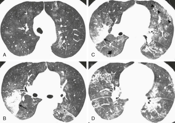
Respiratory syncytial virus pneumonia: chest CT findings.
A–D, Axial chest CT displayed in lung windows shows multifocal, bilateral patchy areas of ground-glass opacity associated with more focal right upper lobe posterior segmental consolidation (arrows). In some areas the ground-glass opacity is associated with intralobular interstitial thickening and interlobular septal thickening (arrowheads). Small solid nodules (C,double arrowheads) are also present.
(Courtesy Michael Gotway, MD.)
