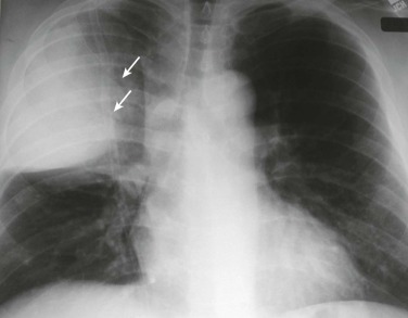
eFigure 33-1.

Lobar pneumonia due to pneumococcus.
Frontal chest radiograph shows homogeneous increased opacity conforming to the shape of the right upper lobe, extending to the pleural surfaces, associated with air bronchograms (arrows). These findings are typical of air space consolidation, and the pattern is consistent with lobar pneumonia, commonly seen with pneumococcal or Klebsiella pulmonary infections.
(Courtesy Michael Gotway, MD.)
