eFigure 33-4.
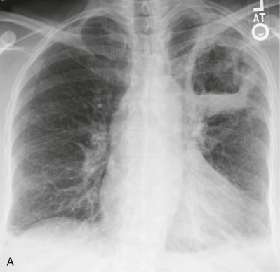
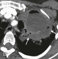
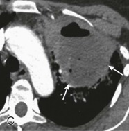
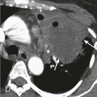
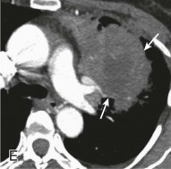
Lung abscess.
A, Frontal chest radiograph in a patient with cough and purulent sputum shows a thick-walled cavity with an irregular internal lining and air-fluid level in the left apex. B–E, Axial chest CT displayed in soft tissue windows shows a rounded area of low attenuation (arrows) in the left upper lobe, surrounded by consolidation consistent with a pulmonary abscess. The internal low attenuation is fluid density, and an air-fluid level is present, typical of pulmonary abscess. Reactive prevascular lymph node enlargement is also evident.
(Courtesy Michael Gotway, MD.)
