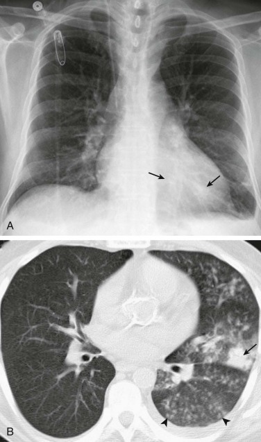
eFigure 33-6.

Pneumococcal bronchopneumonia.
A, Frontal chest radiograph shows patchy bronchovascular thickening (arrows) in the left lower lobe; trace blunting of the left costophrenic angle is present. B, Axial chest CT 2 days following A shows nodular lingular consolidation (arrow) and numerous small centrilobular nodules (arrowheads) consistent with bronchopneumonia. This bronchopneumonia pattern contrasts with the lobar pneumonia pattern (see Fig. 33-1 and eFig. 33-1). Both imaging patterns may be seen with pneumococcal pneumonia.
(Courtesy Michael Gotway, MD.)
