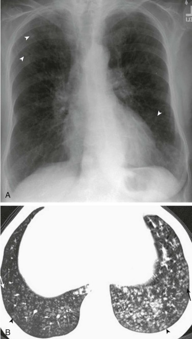
eFigure 33-8.

Haemophilus influenzae pneumonia: bronchiolitis.
A, Frontal chest radiograph shows several nonspecific small nodular opacities bilaterally (arrowheads). B, Axial chest CT through the lung bases shows numerous small centrilobular nodules (arrowheads), some with branching configurations (arrows), the latter consistent with “tree-in-bud” opacity, representing infectious bronchiolitis. The appearance of small centrilobular nodules with branching configurations is not specific for Haemophilus influenzae pneumonia and can be seen with other bacteria and occasionally with fungi or even viruses.
(Courtesy Michael Gotway, MD.)
