eFigure 33-15.

Pseudomonas aeruginosa cavitary pneumonia.
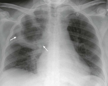
Frontal chest radiograph in a patient with P. aeruginosa pneumonia shows a large right upper lobe thick-walled cavity (arrows).
(Courtesy Michael Gotway, MD.)

Pseudomonas aeruginosa cavitary pneumonia.
Frontal chest radiograph in a patient with P. aeruginosa pneumonia shows a large right upper lobe thick-walled cavity (arrows).
(Courtesy Michael Gotway, MD.)