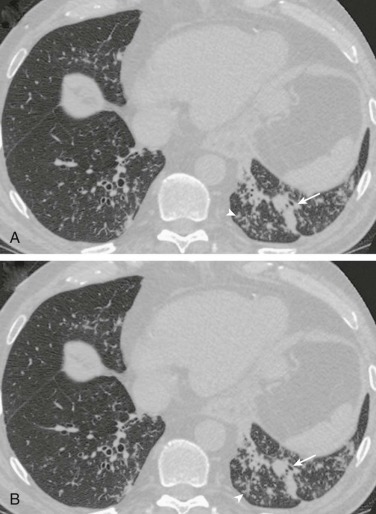
eFigure 33-18.

Aspiration pneumonia: bronchopneumonia/bronchiolitis appearance at chest CT.
A and B, Axial chest CT through the lower lobes displayed in lung windows in a patient with swallowing dysfunction shows numerous small centrilobular nodules, some with branching morphologies (arrowheads), and peribronchial consolidation (arrows). The imaging appearance is consistent with bronchopneumonia, but not specific for aspiration. Note resemblance of this CT appearance with P. aeruginosa pneumonia (see eFig. 33-14), Haemophilus influenzae pneumonia (see eFig. 33-8B), and pneumococcal bronchopneumonia (see eFig. 33-6).
(Courtesy Michael Gotway, MD.)
