eFigure 33-21.
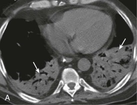
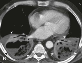
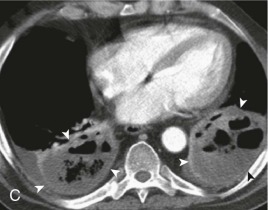
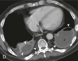
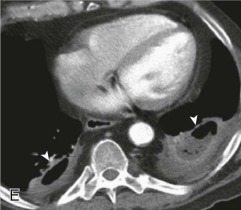
Evolution of pulmonary aspiration into lung abscess.
A, Axial chest CT displayed in soft tissue windows performed shortly following a witnessed aspiration event shows extensive bilateral lower lobe consolidation (arrows). B, Contrast-enhanced chest CT performed several days following A shows developing necrosis and cavitation (arrowheads) within the lower lobe consolidation. C and D, Repeat contrast-enhanced chest CT performed over the ensuing week following A and B shows maturation of frank bilateral lower lobe pulmonary abscesses (arrowheads); note well-defined, enhancing walls surrounding these gas and fluid collections. E, Axial enhanced chest CT following 3 weeks of antibiotic therapy shows partial resolution of the bilateral lower lobe pulmonary abscesses (arrowheads).
(Courtesy Michael Gotway, MD.)
