eFigure 33-22.
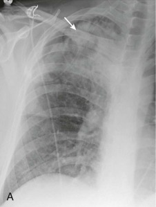
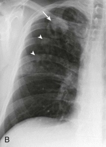
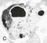
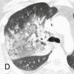
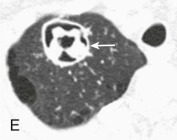
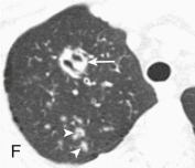
Actinomycosis: cavitary nodule.
A, Frontal chest radiograph shows right upper lobe consolidation and a poorly defined nodular opacity (arrow). B, Frontal chest radiograph 2 weeks following A shows resolution of the right upper lobe consolidation, now exposing a dominant cavitary right apical nodule with internal opacity (arrow), and small surrounding nodules (arrowheads). C and D, Axial chest CT displayed in lung windows performed within 1 day of the presenting chest radiograph (A) shows the right apical opacity as a cavitary nodule with an internal air-fluid level; surrounding ground-glass opacity and consolidation are present, as seen on the chest radiograph (A). E and F, Chest CT displayed in lung windows performed the same day as B shows the dominant right apical opacity with complex internal architecture (arrows) and confirms small surrounding nodules. Biopsy of this lesion recovered Actinomyces israelii.
(Courtesy Michael Gotway, MD.)
