
Fig. 21.41.
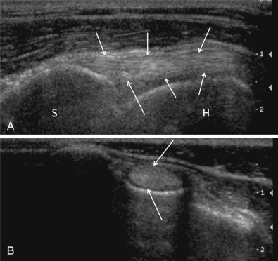
Composite image showing longitudinal (A) and cross-sectional (B) images of the normal biceps tendon (arrows). In a longitudinal plane (A), the tendon is characterized by a linear hyperechoic fiber pattern. The tendon arises from the supraglenoid tubercle of the scapula (S) and extends distally within the bicipital groove along the medial border of the humerus (H). In cross section (B), the tendon is a uniformly hyperechoic oval structure surrounded by a thin hypoechoic rim representing slight fluid in the tendon sheath.
(Reproduced with permission from Davies S, Allan G, Nicoll R: Joints—general. In Kirberger R, McEvoy F, Editors: BSAVA manual of canine and feline musculoskeletal imaging, ed 2, Gloucester, 2016, BSAVA.).