
FIGURE 134-5.
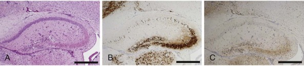
Reovirus-induced apoptosis in the murine central nervous system.
Consecutive sections of the hippocampus prepared from a newborn mouse 10 days following intracranial inoculation with reovirus strain type 3 Dearing. Cells were stained with (A) hematoxylin and eosin, (B) reovirus antigen, and (C) the activated form of apoptosis protease caspase-3. Cells that stain positive for reovirus antigen or activated caspase 3 contain a dark precipitate in the cytoplasm, including neuronal processes. Scale bars, 100 µm.
(Modified from Danthi P, Coffey CM, Parker JS, et al. Independent regulation of reovirus membrane penetration and apoptosis by the µ1 Φ domain. PLoS Pathog. 2008;4:e1000248.)