
Fig. 132.1.
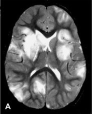
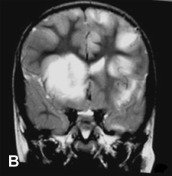
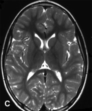
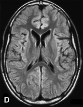
A previously healthy, developmentally normal, 3-year-old boy presented at a local emergency department after 48 hours of emesis, intermittent fever, and lethargy, 1 week after an upper respiratory viral infection. The subsequent neurological involvement included general weakness, drooling, and inability to walk, and he required admission to hospital. One day after admission, the patient presented left-sided hemiparesis followed by status epilepticus. A brain MRI was performed revealing widespread, hyperintense lesions in central and juxta-cortical white matter (A, axial T2-weighted image). Tumefactive lesions involving deep gray and white matter on the right with mass effect were also identified (B, coronal T2-weighted image). ADEM was diagnosed based on clinical and MRI picture and CSF studies including negative viral cultures and viral PCR assays. The child showed rapid improvement following intravenous corticosteroid treatment. Follow-up MRI performed at 6 months, 1 year, and 5 years (C, axial T2-weighted image; and D, axial FLAIR image) after the acute event revealed complete resolution of demyelinating lesions.