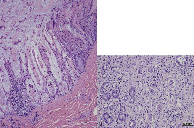
FIGURE 14-5.

A, Segmental crypt necrosis in the ileum of a dog infected with a CPV-2 variant. There is severe loss of crypt epithelial cells. Hematoxylin and eosin (H&E) stain. B, Jejunum of a dog after infection by a CPV-2 variant. Regenerating epithelial cells, which here are nested in an inflamed jejunal lamina, have a large and bizarre appearance and resemble adenoma cells. As a result, the disease has been termed adenomatosis. Parvovirus antigen was no longer detectable by immunohistochemistry at this stage. The crypts in the bottom left corner have a more normal appearance. H&E stain.
(Courtesy Dr. Patricia Pesavento, University of California, Davis Veterinary Anatomic Pathology Service.)
