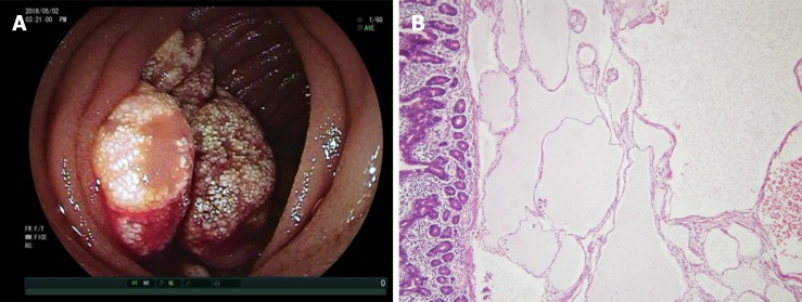
Figure 1.
Gross and histologic images of hemolymphangioma. A: Lobulated tumor occupied half of the intestinal cavity with white patches on the mucosal surface and blood oozing in the fundus; B: Histology revealed a hyperplastic thin-walled lymphangion and venous with luminal dilation in the submucosal area. Hematoxylin and eosin × 20.

