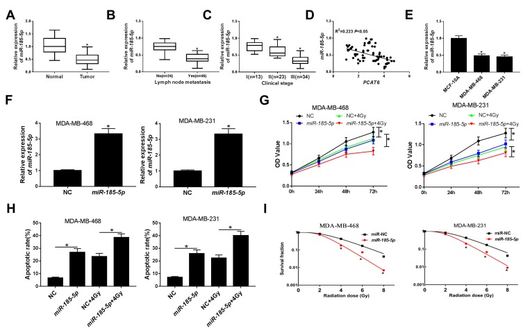
Figure 4.
Effect of miR-185-5p on radiosensitivity of TNBC cells. (A) The expression of miR-185-5p was examined by qRT-PCR in TNBC tissues and matched adjacent normal tissues. (B) The level of miR-185-5p was assessed in tissues from TNBC patients with or without metastasis. (C) The level of miR-185-5p was analyzed in tissues from patients at different clinical stages. (D) The association between PCAT6 level and miR-185-5p abundance was measured in TNBC tissues. (E) The level of miR-185-5p was analyzed in TNBC cells (MDA-MB-468 and MDA-MB-231) and breast epithelial cells (MCF-10A). (F) The transfection efficiency of miR-185-5p mimics was confirmed by qRT-PCR in MDA-MB-468 and MDA-MB-231 cells. (G) Cell viability was evaluated by CCK-8 assay in MDA-MB-468 and MDA-MB-231 cells transfected with NC or miR-185-5p and/or treated with 4Gy irradiation. (H) Flow cytometry analysis was applied to measure the cell apoptosis rate in MDA-MB-468 and MDA-MB-231 cells transfected with NC or miR-185-5p and/or treated with 4Gy irradiation. (I) Colony formation assay was used to determine colony survival fraction in MDA-MB-468 and MDA-MB-231 cells transfected NC or miR-185-5p and treated with different doses of irradiation. *P<0.05.

