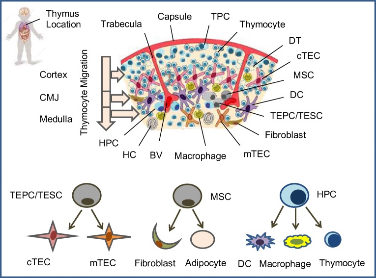
Fig. 1.
Human thymus cell architecture. The human thymus is located in the upper anterior part of the chest behind the sternum between lungs and lies on top of the heart along the trachea. The thymus reaches its maximum weight (about 28 gram) during puberty. This pinkish-gray organ consists of two lobes parted into lobules by connective tissue strands (trabeculae). Each thymic lobule has a cortex and medulla. Hematopoietic precursor cells (HPC) enters the thymus through postcapillary venules located at the corticomedullary junction (CMJ) and migrate to the capsule, committed CD4-CD8- T precursor cells (TPC) located in the subcapsular region, and immature CD4+CD8+ cortical thymocytes migrate through the cortex and CMJ to the medullar zone. The medulla contains CD4+ and CD8+ naïve thymocytes that will migrate to the periphery. The stromal-epithelial compartment of the thymus is represented by minor populations of EpCam+(CD326+)Foxn1+ bipotent thymic epithelial precursor cells/thymic epithelial stem cells (TEPC/TESC) and mesenchymal stem cells (MSC) located probably in the thymic parenchyma close to the CMJ region, as well as EpCam+CD205+ cortical thymic epithelial cells (cTEC) located in the cortex and EpCam+Air+ medullary thymic epithelial cells (mTEC) located in the medulla. Moreover, the cortex and the medulla contain also macrophages, fibroblasts and dendritic cells (DC) that together with cTEC and mTEC participate in the differentiation, maturation, positive and negative selection of thymocytes. HPC generate all thymocyte populations and alternatively may generate macrophages and DC; TEPC/TESC generate cTEC and mTEC lineages depending on local microenvironment and cross-talk with cortical or medullary thymocytes; MSC generate thymic fibroblasts and adipocytes. BV: Blood vessel; DT: Dead thymocyte; HC: Hassall’s corpuscle.

