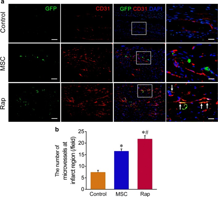
Fig. 6.
Regeneration of the microvessels after cell transplantation. a The microvessels at the infarct region. In rapamycin group, some of GFP+ cells express CD31, which are located on the wall of the microvessels (arrows). The panels of the fourth row are magnification of the boxes in the panels of the third row. Scale bar = 100 μm (the panels of the first to third rows) and 20 μm (the panels of the fourth row). b Statistic result of the number of the microvessels at the infarct region. n = 6. *p < 0.01 versus control group; #p < 0.01 versus MSC group

