Figure 1.

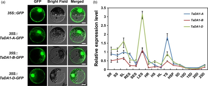
Characterization of TaDA1 homoeologs. (a) Subcellular localization of TaDA1 homoeologs in wheat protoplasts. GFP and TaDA1s‐GFP fusions under the control of the CaMV 35S promoter were transiently expressed in wheat protoplasts. Bar = 10 µm. (b) Tissue expression patterns of the three homoeologs in various Chinese Spring tissues. SR, seedling roots; SS, seedling stems; SL, seedling leaves; RES, roots at elongation stage; SES, stems at elongation stage; LES, leaves at elongation stage; HR, roots at heading stage; HS, stems at heading stage; HL, leaves at heading stage; YS, 1–2‐cm young spikes; HSP, spikes at heading stage; 5D–25D, grains at 5–25 days post‐anthesis, respectively. Normalized values of TaDA1 expression relative to Actin were given as mean ± SD from three replicates.
