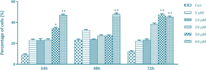
Fig. 2:
SFN enhanced apoptosis in breast cancer cells. Human breast cancer MDA-MB 231 cells were treated with increasing concentrations of SFN (0–40 μM) for 24, 48 and 72 h. The flow cytometry assay was used to assess cell viability and the results were normalized to the untreated control cells. Quantitative data show a significant increase of apoptosis at 30 and 40 μM of SFN in compare with control cells. * and ** donate P<0.05 and P<0.01 respectively, as compared with the control

