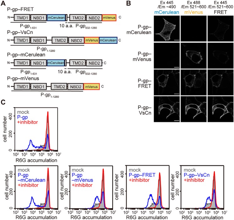
Figure 1.
Localization and function of fluorescent protein-tagged P-gp constructs. A, schematic representations of the fluorescent protein-tagged P-gps. a.a., amino acid. B, localization of the fluorescent protein-tagged P-gp constructs. Each P-gp construct was transiently expressed in HEK293 cells. Fluorescent images were obtained at 37 °C using an LSM700 confocal microscope (Zeiss). Scale bars, 10 μm. C, substrate transport activity. HEK293 cells transiently expressing each P-gp construct were incubated with 1 μm R6G with or without 25 μm inhibitor (PSC-833) for 30 min at 37 °C. R6G accumulation in the cells was measured by flow cytometry.

