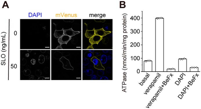
Figure 5.

membrane permeabilized semi-intact cells. A, DAPI staining of cells expressing P-gp–mVenus permeabilized with 50 ng/ml SLO. Fluorescent images were obtained at 37 °C on a Nikon C2 confocal system equipped with a Plan Apochromat ×40/dry objective lens. Scale bars, 10 μm. B, effect of DAPI on the ATPase activity of human P-gp. Proteoliposomes were incubated for 30 min at 37 °C with 100 μm verapamil chloride or 2 μg/ml DAPI in the absence or presence of 1 mm beryllium fluoride (BeFx, an ATPase inhibitor). The values represent the means ± S.D. from three technical replicates.
