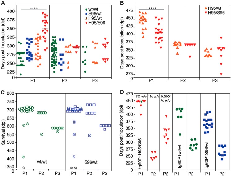
Figure 5.
Selection, adaptation, and unstable propagation of CWD prion lineages in tg33 and tg60 mice expressing deer G96 (WT) and Ser-96–PrPC, respectively. Unless otherwise noted, all inocula were 10% w/v brain homogenates. A, incubation periods for tg33 mice infected with WT/WT (green), Ser-96/WT (blue), His-95/WT (orange), and His-95/Ser-96 (red) CWD and their descendent prion conformations. Incubation periods were shortened during serial passage (P1–P3) (****, p < 0.0001; Kruskal-Wallis, Dunn's multiple comparison test). B, rapid adaptation of a prion species (H95+) descending from Wisc-1 infection in His-95/WT and His-95/Ser-96 deer during passage in tg60 mice (****, p < 0.0001; Mann-Whitney test). C, unstable propagation of WT/WT and Ser-96/WT prions in tg60 mice failed to produce clinical prion disease. Open symbols represent mice that survived until the end of the experiment. Crossed symbols represent mice with intercurrent disease or time points for PrPCWD evaluation and subsequent passage. D, comparison of prion doses accumulated in tg60 mice by bioassay in tg33 mice. P1 inocula are indicated and were from tg60 mice euthanized after 359 (red; shown in B) and 345 dpi (crossed symbols; shown in C). Stable H95+ prion replication with Ser-96–PrPC generated ∼10-fold more prions than WT/WT and Ser-96/WT prion species. Some transmission data (P1, except for D, and P2 for the tg60 in B) were previously reported (24) and are reproduced here for comparison. A single modification was made to the reproduced data (C; time points 345 days post inoculation were not previously reported). The P2 in tg60 mice in B was also reproduced from Ref. 24.

