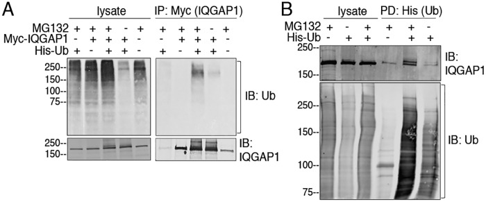
Figure 1.
Ubiquitination of IQGAP1. A, HEK293 cells were transfected with (+) or without (−) Myc-IQGAP1 and/or His-ubiquitin (Ub). Where indicated, cells were incubated with MG132 (+) or DMSO (−) (vehicle) for 4 h. Equal amounts of protein lysate were loaded directly onto gels or immunoprecipitated (IP) with anti-Myc beads. Proteins were analyzed by SDS-PAGE and immunoblotting (IB) using anti-ubiquitin and anti-IQGAP1 antibodies. B, HEK293 cells were transfected with (+) or without (−) His-ubiquitin and incubated with MG132 (+) or DMSO (−). His-ubiquitin was pulled-down (PD) by incubating equal amounts of protein lysate with TALON beads. Lysates and complexes were resolved by Western blotting. The PVDF membranes were probed with anti-ubiquitin and anti-IQGAP1 antibodies. All data are representative of at least three independent experiments.

