Figure 1.

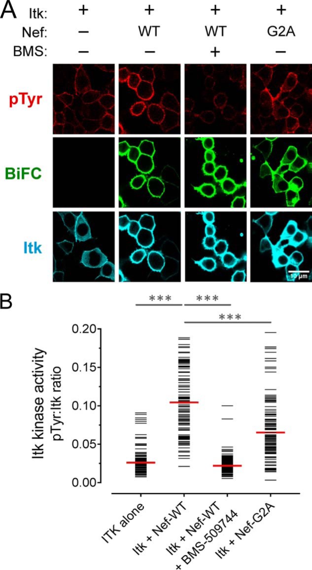
Nef induces constitutive activation of Itk at the cell membrane. A, Itk was expressed either alone or together with WT or myristoylation-defective Nef (G2A) as BiFC pairs in the absence or presence of the Itk inhibitor BMS-509744 (1 μm) in 293T cells. The cells were fixed and stained for confocal microscopy with anti-pTyr antibodies as a measure of kinase activity (red) and anti-V5 antibodies to verify Itk protein expression (blue). Nef interaction with Itk is observed as fluorescence complementation of the YFP variant, Venus (BiFC; green). B, single-cell image analysis. Mean fluorescence intensities for the pTyr and Itk signals were determined for ≥100 cells from each condition using ImageJ. The fluorescence intensity ratio (pTyr:Itk expression) for each cell is shown as a horizontal bar, with the median value indicated by the red bar. Student's t tests were performed on the groups indicated by horizontal lines above the plot; p < 0.0001 in each case (***).
