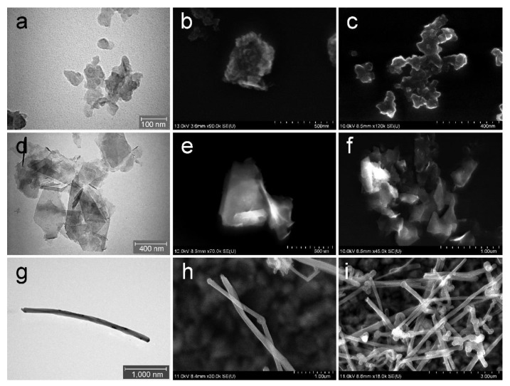
Figure 1.
Characterisation of C-BNM by electron microscopy: (a) TEM and (b,c) SEM detail of the GP1 forming small aggregates; (d) TEM detail of the GP2; (e) SEM detail of a structure of GP2 single platelet and (f) forming clusters; (g) TEM detail of single MWCNT; (h) SEM detail of a structure of MWCNT with (i) forming clusters.

