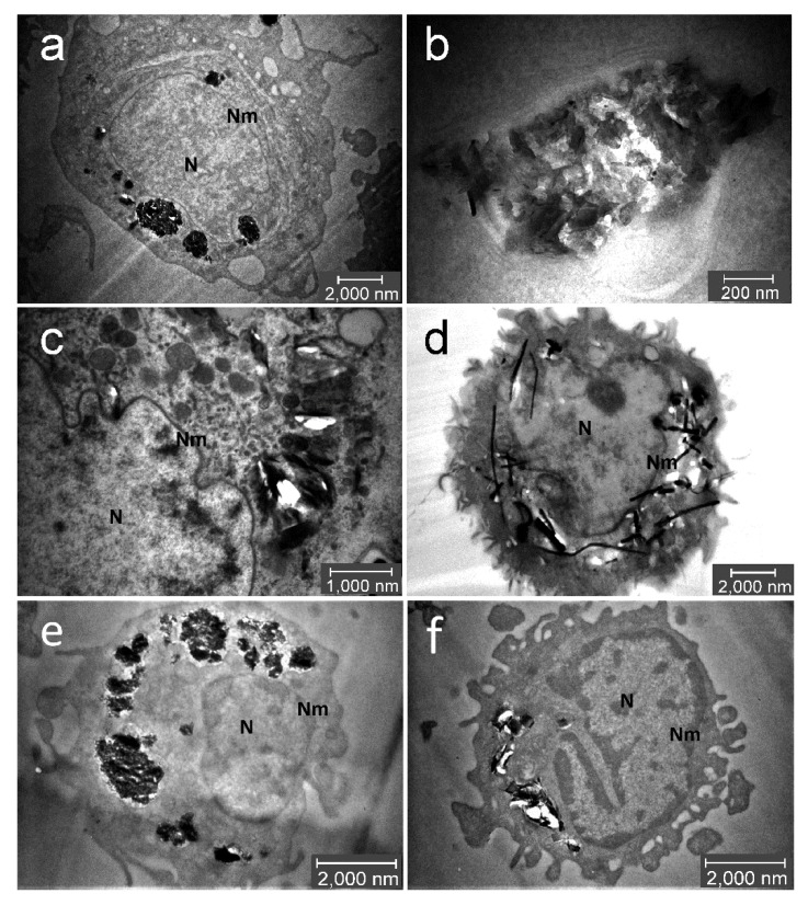
Figure 4.
Intracellular localisation of C-BNM after 24 h exposure in THP-1 (a–d) and primary monocytes (e,f): GP1 (a,b,e) forms large aggregates in cytoplasm apparently in vesicles. No particles are found in nucleus (N); (c,f) GP2 forms smaller aggregates in cytoplasm, apparently in vesicles. Occasionally, free particles are detected in cytoplasm; (d) MWCNT were found as free needle-like objects in the cytoplasm, possibly from damaged lysosomes. Sporadically, they can be found in nucleus or penetrating through the nuclear membrane (Nm).

