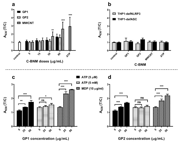
Figure 6.
Effect of C-BNM on activation of inflammasome NLRP3 after 24 h exposure: (a) NLRP3 activation in THP1 null cells was measured as conversion of proIL-1β to IL-1β, which was detected using HEK-Blue™ IL-1β cells. Data were normalised to the control (untreated THP1-null cells). ATP was used as positive standard of NLRP3 induction. The symbols ** p < 0.01; *** p < 0.001 highlight the statistical significance as compared to the corresponding control; (b) Activation of NLRP3 in deficient cells THP1-defNLRP3 and THP1-defASC, which were treated the same way as THP1-null cells; (c,d) activation of NLRP3 in THP-1 null macrophages by GP in presence of MDP and ATP: ATP (5 mM; 5 µM) and MDP (10 µg/mL) were used as a standard activators of NLRP3 (control) in presence of 0, 25 and 50 µg/mL of GP. The symbols * p < 0.05; ** p < 0.01; *** p < 0.001 highlight statistical significance as compared to the corresponding controls (0) without GP.

