Figure 7.
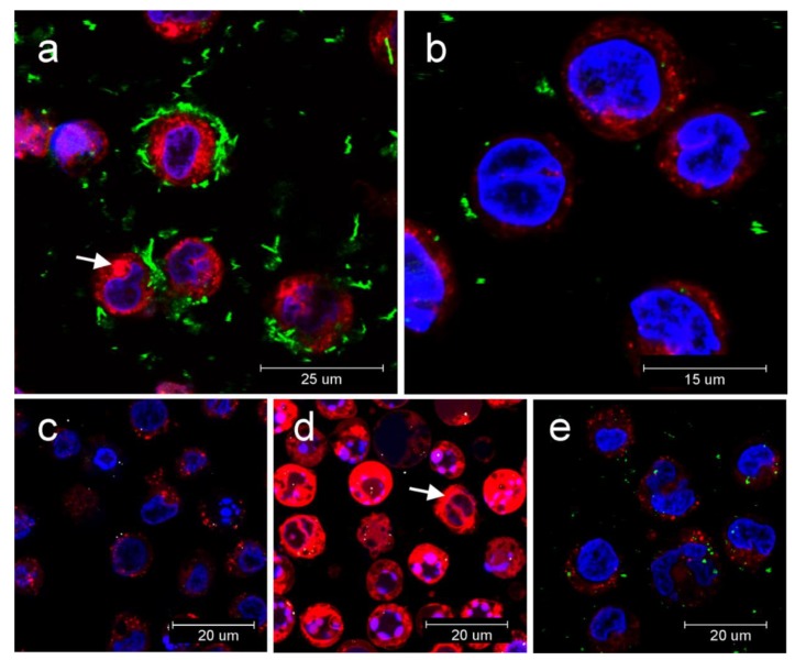
Release of cathepsin B from lysosomes into cytoplasm in THP1-null cell revealed by confocal microscopy: Proteolytic activity of cathepsin B was determined by fluorogenic substrate with red emission; (a) release of cathepsin B (red fluorescence) into cytoplasm after 24 h incubation with MWCNT (light scattering in green). Cytoplasm stained with fluorogenic substrate (white arrow); (b) Release of cathepsin B after 24 h incubation with GP2 (light scattering in green); (c) Negative control; (d) release of cathepsin B after incubation with lysosomal disruptor LLME with burst of cathepsin B into cytoplasm (white arrow); (e) release of cathepsin B after 24h incubation with GP1 (light scattering in green).

