Figure 3.
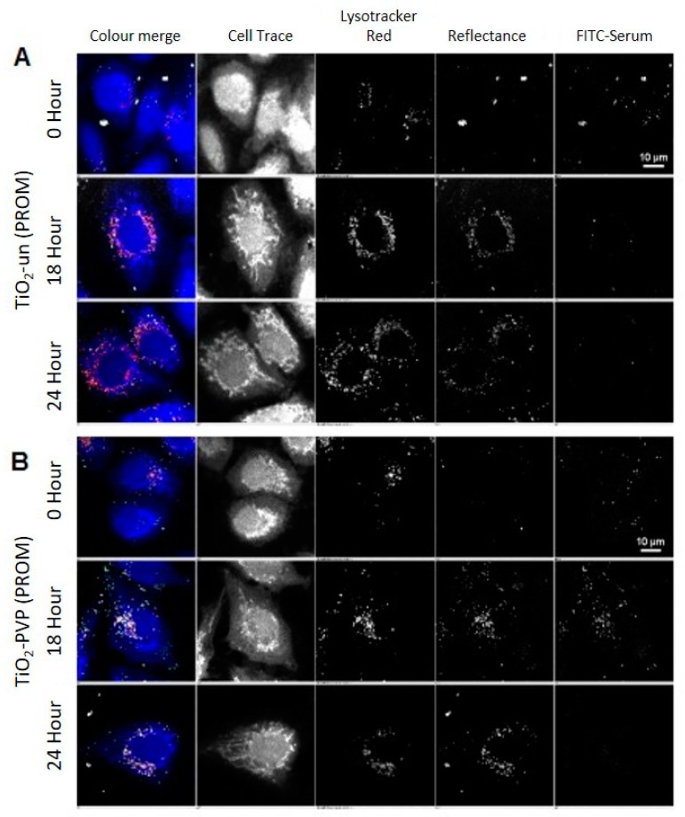
Confocal acquisition of A549 cells treated with uncoated (TiO2-un) and PVP-coated (TiO2-PVP) TiO2 NMs compared to the control (untreated cells) in FITC-labeled serum, followed by post-acquisition in unlabeled serum and fixation at 15 min (0 h), 2, 6, 18, and 24 h post-exposure. (A) Images show the presence of TiO2-un as large, bright agglomerates associated with the surface of the cell, which are subsequently internalized and trafficked to lysosomes marked by lysotracker red. Past the 0 h time point, TiO2-un rapidly loses the FITC signal, suggesting that in the absence of a surface coating, the protein corona is not stably bound. (B) TiO2-PVP, on the other hand, demonstrates an increase of the FITC signal up to the 18 h time point, after which there is a significant loss of signal. As NMs co-localize well with lysotracker at 18 and 24 h, this is likely due to degradation of the protein corona within the acidic lysosomal compartment.

