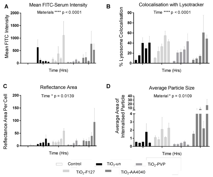
Figure 5.
(A) Quantification of confocal data at 0, 2, 6, and 24 h post-exposure shows a change in the FITC-serum signal over time. While TiO2-un NMs rapidly lose the FITC-serum signal, surface-modified particles retain this, with a steady increase over time. TiO2-PVP demonstrates a slight reduction in signal between 18 and 24 h, potentially indicative of lysosomal degradation. (B) All four NMs demonstrate a steady increase in lysosomal co-localization, with significance demonstrated over time (**** p < 0.0001), but not between materials. (C) An increase in the reflectance area over time, indicative of NM uptake, is observed (* p = 0.0139), with no significant difference in NMs observed, consistent with the equivalent exposure of each NM type to the cells in question. (D) Finally, a difference in NM agglomerate size (in microns) is noted between materials (* p = 0.0109) consistent with images which show TiO2-PVP internalized as small discrete agglomerates, compared to the larger agglomerates of TiO2-F127 and TiO2-AA4040 observed (two-way ANOVA, SEM, n = 3).

