Figure 1.

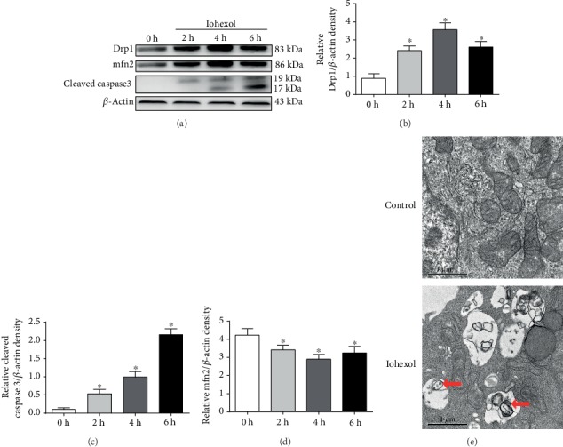
Iohexol induces cytotoxicity and mitochondrial dysfunction in HK-2 cells. HK-2 cells were treated with iohexol (200 mg I/ml) at indicated time points. (a) Drp1, mfn2, and cleaved caspase 3 were measured by Western blot assay (n = 3). Iohexol treatment caused a decreased expression of mfn2 and increased expression of Drp1 and cleaved caspase 3. (b–d) Quantification of average Western blot band intensities. (e) Representative images. HK-2 cells were treated with iohexol (200 mg I/ml) for 4 hours and then collected for transmission electron microscope analysis. Mitophagy was labeled with red arrow. Values were presented as mean ± SE. ∗p < 0.05, compared with the control group.
